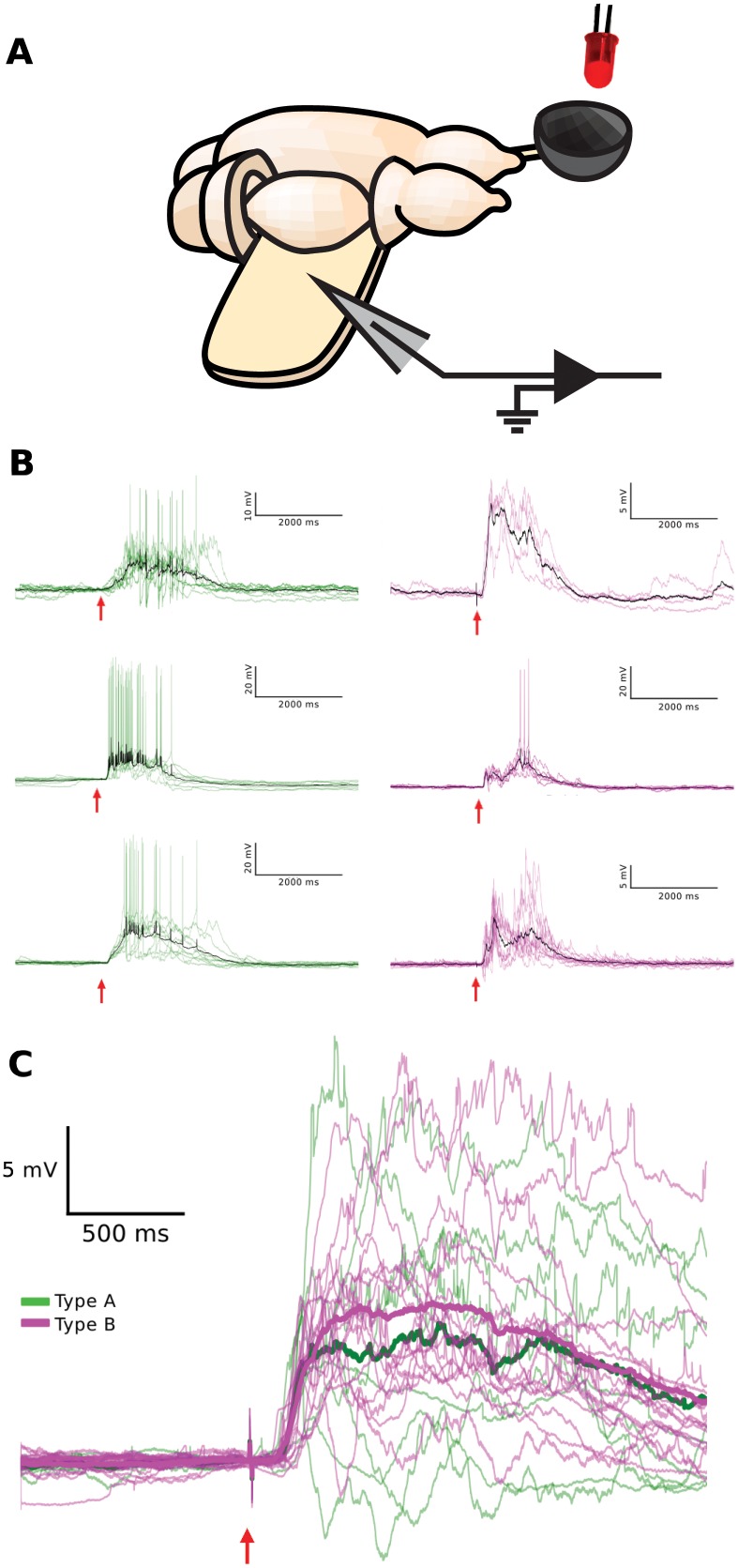
Fig 8. Visual response properties of the two physiologically defined pyramidal neuron types.
(A) Schematic of the turtle ex vivo eye-attached whole-brain preparation. A diffuse flash of light from the LED (red) is projected onto the intact retina within the eye-cup (gray bowl), while the membrane potential from a pyramidal neuron is recorded with a patch electrode (gray triangle) inserted into the unfolded visual cortex. (B) Pyramidal neuron membrane potential responses to brief flashes of light (10 ms, 640 nm; red arrow) persist long beyond the duration of the flash, are variable from trial-to-trial, and display sparse spiking. Trial averages are shown in black. Representative membrane potential visual responses to flashes are shown for three pyramidal neurons from each physiologically defined type (green/A and magenta/B). The responses are fluctuating and similar for both types. (C) The time courses of trial-averaged membrane potential (after spike clipping) of all pyramidal neurons recorded in response to flashes (8 type A (green), 16 type B (magenta)). Averages across pyramidal neuron visual responses of the same physiological type are plotted in bold (green/A and magenta/B).

