Fig. 2.
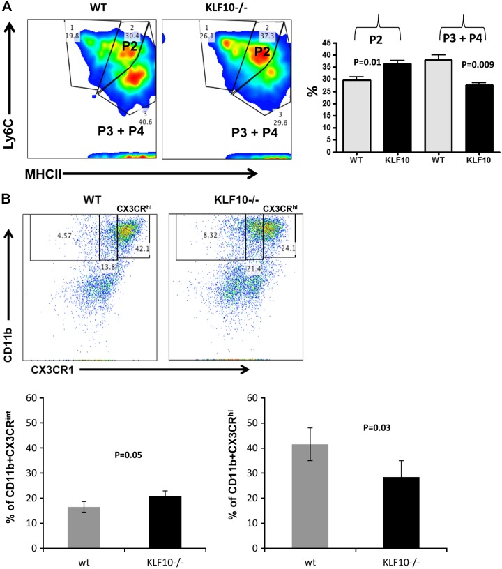
Characterization of colonic macrophages in adult (6-to 8-wk-old) KLF10−/− mice. A: Lamina propria mononuclear cells (LPMC) were isolated from WT or KLF10−/− mice and stained for CD45, CD11b, CD64, CD103, LyC6, and MHCII and analyzed by flow cytometry. Cells were gated on CD45+ live CD11b+CD64+CD103− subset and analyzed for the expression of Ly6C and MHCII. The different macrophage subsets were then characterized as proinflammatory (Ly6C+MHCII+) P2 or anti-inflammatory (Ly6C−MHCII+) P3 and P4. Average percentages ± SE of the different colonic macrophage subsets P2, P3, and P4 in WT and KLF10−/− colon are shown (right, n = 4). B: representative dot-plot analysis of CX3CR1 expression among CD11b+ murine colonic mononuclear cells between WT and KLF10−/− mice at day 8 following DSS treatment. Bottom: average percentages of CX3CR1int or CX3CR1hi subset in WT vs. KLF10−/− mice (n = 4 mice/group).

