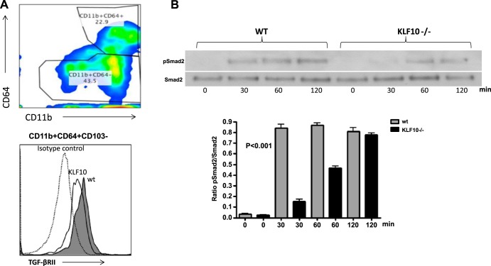
Fig. 4.
Colonic macrophages in KLF10−/− mice express lower levels of TGFβRII and have altered TGF-β signaling. A: colonic macrophages were isolated and stained for CD45, CD11b, CD64, and CD103. Cells were gated on CD11b+CD64+ cells and analyzed for the expression of TGFβRII. The depicted representative histogram indicates the MFI of TGFβRII expression in WT vs. KLF10−/− colonic macrophages from 3 independent experiments with similar results. B: attenuated early Smad-2 phosphorylation in KLF10−/− macrophages in response to TGF-β1 stimulation. For the analysis of pSmad2, cells were serum starved for 24 h and activated for the indicated time points with TGF-β1 (5 ng/ml) lysed and analyzed for p-Smad2 and total Smad2 by Western blotting. Representative of 4 experiments with similar results is shown. The average ratio of p-Smad2/total Smad2 between WT and KLF10−/− macrophages at the indicated time points from 4 independent experiments is shown at bottom (P < 0.001 by 1-way ANOVA analysis).

