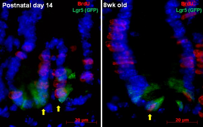
Fig. 5.
Localization of BrdU (red) and Lgr5 (green fluorescent protein, GFP, green) in small intestinal crypt epithelial cells of WT mouse at age 14 days and at age 8 wk. Colocalization of BrdU and Lgr5+ (arrows) identifies proliferating crypt epithelial stem cells. Scale bar = 20 μm, original magnification ×1,000.

