Fig. 6.
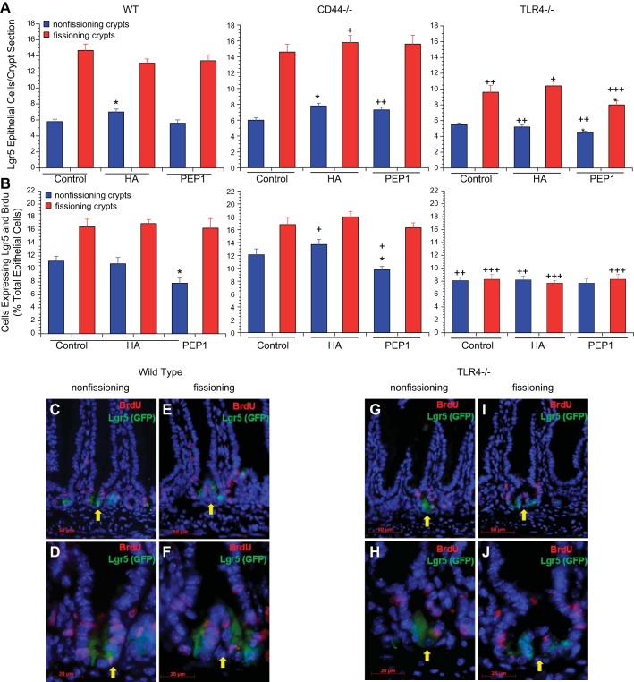
Effect of genotype and treatment with HA or PEP-1 on Lgr5+ cell number and proliferation. A: Lgr5+ cell number. Effect of genotype was measured; 14-day-old WT and CD44−/− mice had similar numbers of Lgr5+ crypt epithelial cells per section in nonfissioning and fissioning crypts. TLR4−/− mice also had a similar number of Lgr5+ epithelial cells at baseline in nonfissioning crypts but had significantly fewer Lgr5+ epithelial cells in fissioning crypts compared with WT or CD44−/− mice. Overall Lgr5+ cell numbers were significantly diminished in 14-day-old TLR4−/− mice compared with WT and CD44−/− mice, especially in fissioning crypts. +P < 0.01, ++P < 0.001, +++P < 0.0001 compared with WT mice given the same treatment. Effect of treatment was measured; postnatal WT and CD44−/− mice showed a small increase in Lgr5+ cell numbers in nonfissioning crypts after treatment with exogenous HA from age 7 days to age 14 days, but there was no increase in TLR4−/− mice. Blocking endogenous HA by intraperitoneal administration of PEP-1 from age 7 days to age 14 days had no effect on Lgr5+ cell numbers in WT or CD44−/− mice, but there was a small further diminishment of Lgr5+ cell numbers in TLR4−/− mice. Values are means ± SE for 6 mice per treatment group. *P < 0.01 compared with control of the same genotype. B: Lgr5+ intestinal crypt epithelial cell proliferation. Effect of genotype was measured, and 14-day-old WT and CD44−/− mice had similar baseline rates of Lgr5+ intestinal crypt epithelial cell proliferation, with higher percentages of Lgr5+ epithelial cells proliferating in fissioning than in nonfissioning crypts. 14-day-old TLR4−/− mice had significantly lower rates of Lgr5+ epithelial cell proliferation compared with WT and CD44−/− mice. TLR4−/− mice also differed from WT and CD44−/− mice in having a rate of Lgr5+ epithelial proliferation that was no greater in fissioning crypts than in the nonfissioning crypts. +P < 0.01, ++P < 0.001, +++P < 0.0001 compared with WT mice given the same treatment. Effect of treatment was measured, and mice were treated with HA or PEP-1 as in Fig. 4. Treatment with HA had no effect on Lgr5+ epithelial cell proliferation in WT, CD44−/−, or TLR4−/− mice. Administration of PEP-1 resulted in a significant decrease in Lgr5+ cell proliferation in nonfissioning intestinal crypts in WT and CD44−/− mice, while not affecting Lgr5+ cell proliferation in fissioning crypts. PEP-1 had no effect on Lgr5+ epithelial cell proliferation in TLR4−/− mice. Values are means ± SE for 6 mice per treatment group. *P < 0.01 compared with control of the same genotype. C–J: immunofluorescence for BrdU (red) + Lgr5 (GFP, green) showing BrdU-labeled and Lgr5+ epithelial cells in nonfissioning and fissioning small intestinal crypts of postnatal day 14 WT (C–F) and TLR4−/− (G–J) mice. Scale bar for C, E, G, I = 50 μm, original magnification ×400. Scale bar for D, F, H, J = 20 μm, original magnification ×1,000.

