Fig. 9.
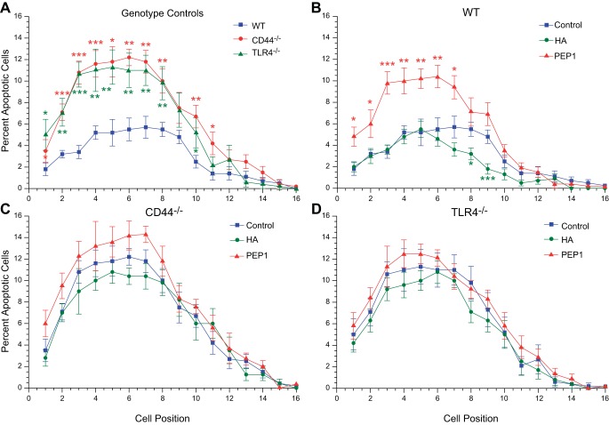
Positional distribution of apoptotic epithelial cells in small intestine of postnatal day 14 WT, CD44−/−, and TLR4−/− mice. A: in all 3 genotype controls, the highest rate of apoptosis was in the range of positions 4–7, with double the rate in CD44−/− and TLR4−/− compared with WT mice. B: intraperitoneal administration of exogenous HA to WT mice from age 7 days to age 14 days reduced apoptosis in positions 7–9 but had no effect below this region of the crypt. Treatment with PEP-1 using the same regimen resulted in a 2-fold increase in apoptosis in the lower half of the crypt (positions 1–7). C and D: exogenous HA and PEP-1 had little to no effect on apoptosis in postnatal CD44−/− and TLR4−/− mice. *P < 0.03, **P < 0.005, ***P < 0.0001 compared with control.

