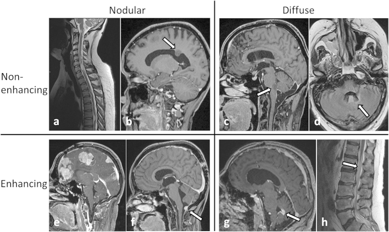
Figure 4. MRI examples of LM in the study population (a–c and e–h: T1w +Gd, d: T2-FLAIRw MRI).
Two general morphological types of LM – nodular (a+b, e+f) or diffuse (c+d, g+h) – were observed. Both types occurred as enhancing (lower row) or non-enhancing (upper row) lesions. White arrows indicate sites of LM.

