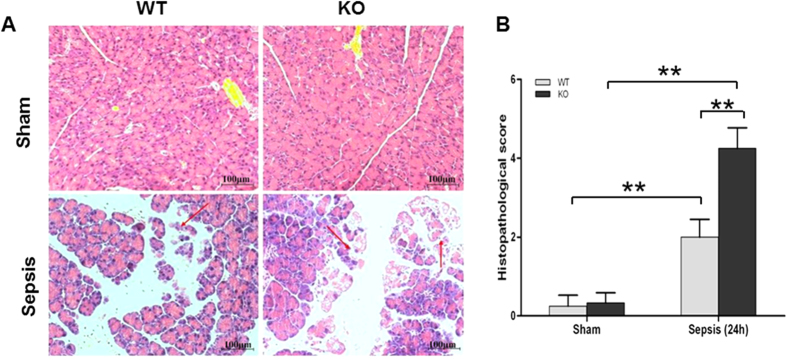
Figure 2. Histopathology of the pancreas in septic WT and KO mice.
Pancreatic tissues from WT mice and SP-D KO mice were examined under the light microscope after staining with H/E (A). The histopathological scores of pancreatic injury were assessed (B). The pancreatic tissues in septic WT and SP-D KO mice showed severe pathological injury 24 h after CLP. Inflammation, edema, and necrosis of the acinar cells were observed. Pancreatic histopathological score index in septic SP-D KO mice was higher than that of septic WT mice (B). magnification × 200. Graphs represent the mean ± SEM. **p < 0.01. scale bars = 100 μm. (n = 6 mice/group).

