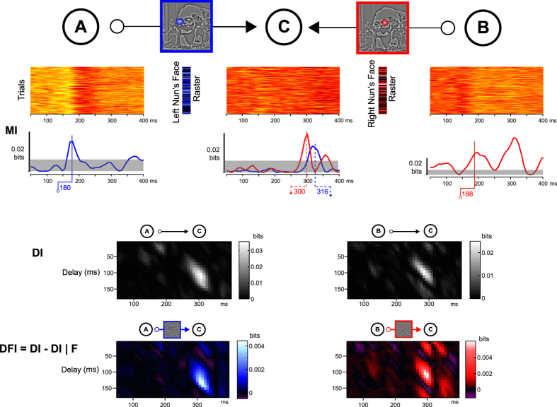
Figure 3. Directed Information (DI) and Directed Feature Information (DFI).
Having identified in Fig. 2 What features the brain of Observer 1 processes for perceptual decisions (framed in blue and red in Fig. 2 and here), with DFI we illustrate coding and transfer of these features between right (node A) and left (node B) occipito-temporal regions and inferior parietal region (node C). MI. Color-coded Mutual Information times courses for each feature (single-trial left and right nun’s faces values, see corresponding color-coded rasters) and network node (single-trial MEG activity). DI. Directed Information reveals that A and B send MEG signal information to C with a ~120 ms of delay. DFI. Directed Feature Information reveals the transfer that is about each feature, reconstructing two links of an information processing network of three nodes. The section of the Dali painting “Slave Market with a Disappearing Bust of Voltaire” is © Salvador Dalí, Fundació Gala-Salvador Dalí, VEGAP, 2014 and is excluded from the Creative Commons license covering the rest of this work.

