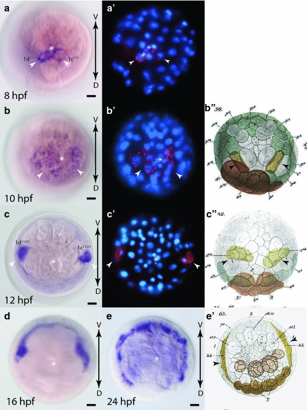
Fig. 10.

Expression of fzCRD-1 during early development in Platynereis. a–e WMISH of fzCRD-1, and a′–c′ false color images of WMISH (red) overlaid with DAPI-stained nuclear images (blue). a, a′ 8 hpf embryo, animal-pole view. White arrowheads indicate 1c112 and 1d112 cells. b, b′ 10 hpf embryo animal-pole view. White arrowheads indicate expression in 1c112 and 1d112 progeny. c, c′ 12 hpf embryo animal-pole view. White arrowheads indicate expression in 1c11221 and 1d11221 which have migrated laterally by this point in development. d 16 hpf and e 24 hpf embryos, animal-pole views showing continued expression in elongating ring canal. b″, c″, e′ Modified images from Wilson (1892) [44] showing migration and elongation of ring canal cells (yellow cells indicated by black arrowheads). WMISH images are shown with ventral at the top to align with Wilson’s original sketches. White asterisks indicate animal pole. Black arrows show the orientation of the dorsal–ventral axis (D-V)
