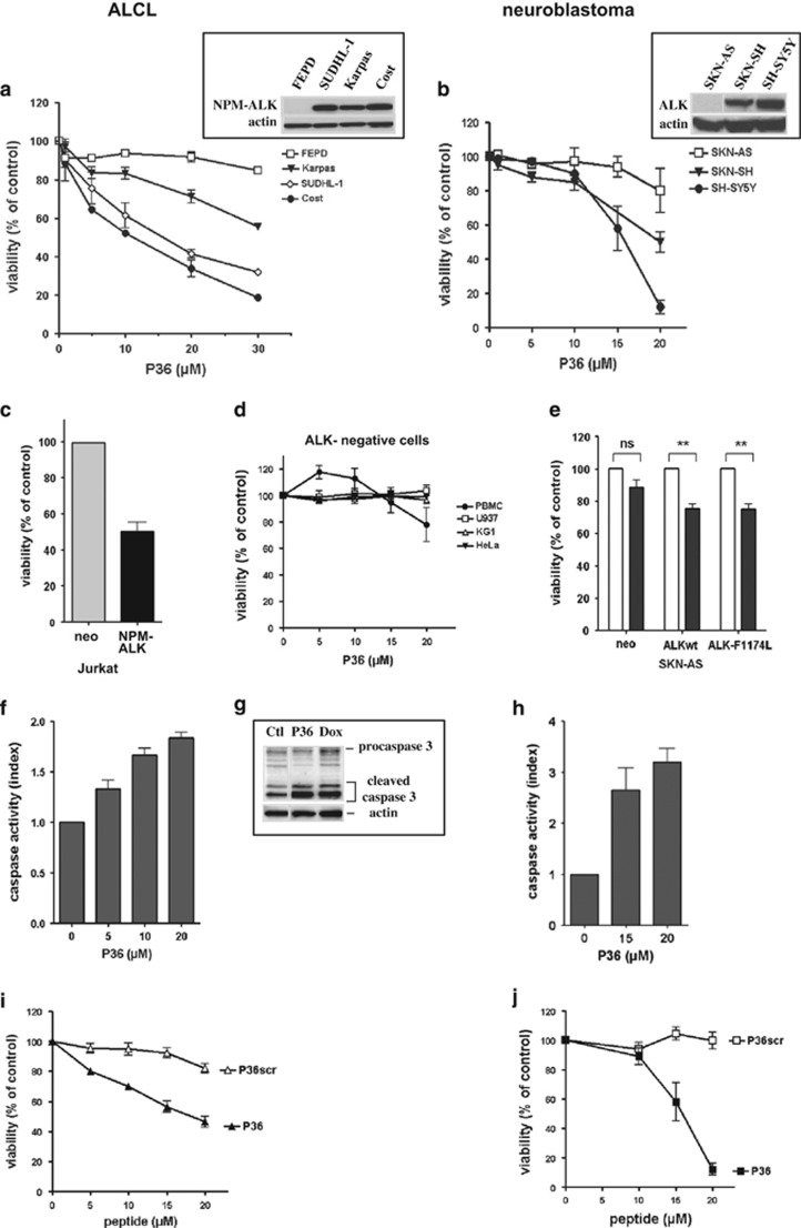
Figure 2.
A peptide (P36) derived from the ADD domain of ALK specifically induces apoptosis in ALK-positive tumor cells. Cells were incubated with various concentrations of P36 for 48 h, or with culture medium without peptide for the control. Top panels: effect of P36 on the viability of ALK-positive and -negative ALCL (a) and neuroblastoma (b) cell lines. Insets: western blot analysis of ALK expression in the cell lines. Actin detection was used as a loading control. (c–e) Specificity of P36-induced apoptosis for ALK-expressing cells. ALK-negative normal (PBMC) or tumor (U937, KG1, HeLa) cells are insensitive to P36 (d). ALK-negative cells stably transfected with ALK variants (NPM–ALK in Jurkat (c), ALKwt or mutant ALK-F1174L in SKN-AS (e)) or an empty vector (neo) control were incubated with 5 μM (Jurkat) or 15 μM (SKN-AS) P36 and their viability assessed at 48 h. (f–h) P36 induces caspase-dependent apoptosis in ALK-positive cells. Cost ALCL (f) or SH-SY5Y neuroblastoma (h) cells were incubated with various doses of P36 for 48 h. Caspase 3/7 activity was measured and expressed as an index relative to control untreated cells (f and h). An increased caspase 3 cleavage was detected by western blot in Cost cells (g) treated for 6 h with P36 (10 μM) or doxorubicin (Dox, 2 μM) compared with culture medium (ctl). (i and j) Specificity of P36 cytotoxic effect on Cost ALCL (i) and SH-SY5Y neuroblastoma (j) compared with a scrambled version (P36scr) of P36. Values in all graphs represent the mean ±S.E.M. from at least three independent experiments. NS, not significant, **P<0.01, statistical test: two-way analysis of variance (using Graphpad Prism 4 software)

