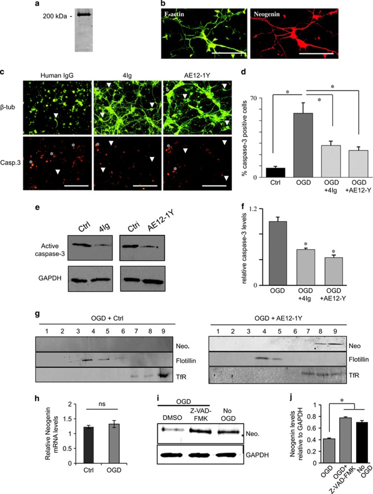
Figure 3.
Blocking Neogenin association with lipid rafts decreases apoptosis and caspase-3 activation. (a and b) Neogenin is expressed in cortical neurons. Primary cortical neurons were cultured and submitted to (a) western blot for Neogenin and (b) double labeling for F-actin (Alexa-Fluor Phalloidin) and Neogenin. Western blot as well as immunofluorescence revealed a strong Neogenin expression. Bar, 100 μm. (c) Primary cortical neurons were subjected to 1 h OGD and then treated with either human IgG (1 μg/ml, control), 4Ig (1 μg/ml) or AE12-1Y (1 μg/ml) for 6 h. Cells were immune stained for active caspase-3 and βIII-tubulin. (d) Quantification of experiments presented in c revealed that OGD resulted in an increase of caspase-3-positive cells (*P<0.005). The addition of both 4Ig and AE12-1Y to the medium significantly decreased the number of caspase-3-positive cells (*P<0.005). Bar, 150 μm. (e) Western blot analysis on cells following OGD plus treatment with control (human IgG), 4Ig and AE12-1Y. The intensity of active caspase-3 bands appeared weaker in the presence of 4Ig and AE12-1Y. (f) Quantification of western blot experiments showed that the presence of 4Ig and AE12-1Y significantly reduced caspase-3 activation (*P<0.005). (g) Cortical neurons were submitted to OGD in the presence of 1 μg/ml of AE12-1Y or control (human IgG 1 μg/ml) and fractionation was performed 1 h after ischemia. In control experiment, Neogenin (Neo.) signal could not be seen. In the presence of AE12-1Y, Neogenin colocalized with the heavy fraction marker transferrin receptor (TfR). Flotillin was used as a marker for lipid rafts. (h) Assessment of the relative Neogenin mRNA levels (relative to HPRT1) in OGD and control cultures revealed no significant (NS) difference. (i) Cortical neurons were treated with the pan caspase inhibitor Z-VAD-FMK (20 μM) or DMSO (control) for 2 h and submitted to OGD for 30 min. Western blotting analysis was performed with Neogenin and GAPDH antibodies. In the presence of Z-VAD-FMK, levels for Neogenin appeared higher. (j) Neogenin levels relative to GAPDH were quantified, which demonstrates that incubation with Z-VAD-FMK restored Neogenin levels. Data from three independent experiments (*P<0.05)

