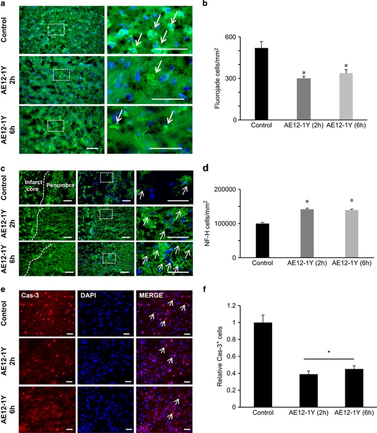
Figure 7.
Treatment with AE12-1Y reduces neuronal damage after MCAO. Animals were submitted to MCAO and treated with human IgG (control) or AE12-1Y at 2 or 6 h following ischemia. One week after insult, brains were sectioned and neuronal damage was assessed. (a) Fluoro-Jade B labeling (green) of degenerating neurons revealed a higher number of damaged neurons in control versus AE12-1Y-treated animals. (b) Quantification indicated that the number of Fluoro-Jade-positive cells was significantly lower in AE12-1Y-treated (2 and 6 h) brains when compared with controls. (c) Fluorescence micrographs of peri-infarct area of the injured hemisphere. Columns (left to right) show NF-200 (NF-H) immunoreactivity with indications of the penumbra and core infarct, a merge of the preceding image with DAPI and a higher magnification view of the inset white box in the merged images. Control samples showed sparse neurofilament immunoreactivity, whereas samples treated with AE12-1Y showed increased levels of NF-200 after MCAO. (d) Quantification of the relative mean number of NF-200-positive cells per mm2 (±S.E.M.) following MCAO. AE12-1Y significantly increased the relative level of NF-200 (*P<0.01), at 7 days after stroke. Scale bar, 100 μm. (e) Representative pictures of control and antibody AE12-1Y treated cortical cultures 2 or 6 h following OGD and staining with DAPI or caspase-3. Bar, 100 μm. (f) Quantification of experiments presented in e showed a significant reduction of caspase-3 activation following AE12-1Y treatment (*P<0.01)

