Figure 2.
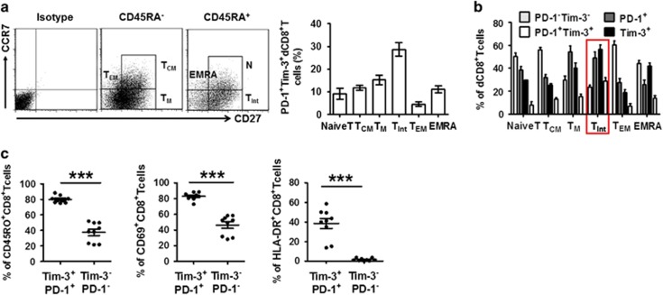
Decidual CD8+ T cells co-expressing Tim-3 and PD-1 display a TInt phenotype. (a and b) Decidual CD8+ T (dCD8+ T) cells from first trimester pregnancy (n=12) were stained with antibodies against CD45RA, CCR7, CD27, or their respective isotype controls to determine their differentiation phenotype (see Results). (a) Frequency of PD-1+Tim-3+ cells within each population. Cells expressing both PD-1 and Tim-3 were predominantly of the TInt phenotype. Data represent mean±S.E.M. The flow-cytometry plot is representative of four independent experiments. (b) Frequency of PD-1–Tim-3–, PD-1+, Tim-3+, and PD-1+Tim-3+ cells within each population. (c) Decidual CD8+ T cells from first trimester pregnancy were stained with antibodies against PD-1, Tim-3, CD69, HLA-DR, and CD45RO. The PD-1+Tim-3+ and PD-1–Tim3– phenotypes were compared. n=9. Data represent mean ±S.E.M. from three independent experiments. A significant portion of PD-1+Tim-3+ T cells expressed all three activation/memory markers. ***P<0.001

