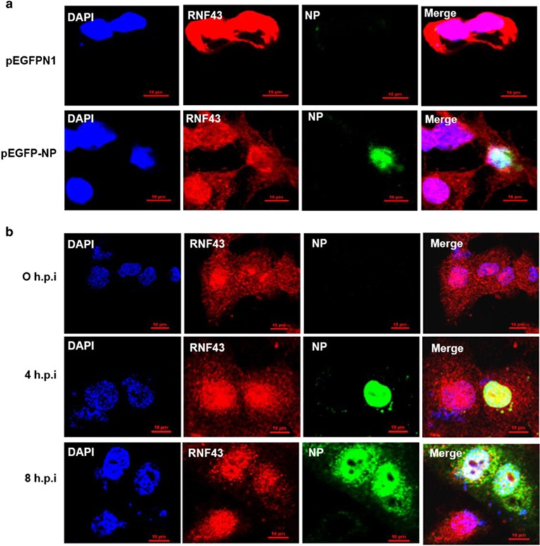
Figure 2.
IAV NP and RNF43 co-localize in the nucleus of mammalian cells. (a) A549 cells were transfected with pEGFPN1 control plasmid or pEGFP-NP. Cells were fixed after 48 h and stained with DAPI for nucleus and anti-goat secondary antibody conjugated to Alexa-594 for RNF43 (red) and observed under confocal microscope. GFP-tagged NP is shown in green. (b) A549 cells were infected with PR8 IAV with 5 MOI and were fixed at the indicated time points. NP was stained using anti-NP monoclonal primary antibody and anti-mouse Alexa488 conjugated secondary antibody (green). RNF43 was stained using specific primary antibody and anti-rabbit Alexa-594 conjugated secondary antibody (red). Panels are labeled for their respective staining, RNF43 (red), NP (green) and nucleus (blue)

