Figure 3.
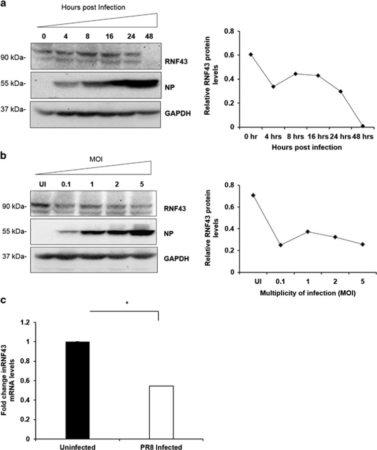
IAV infection decreases abundance of RNF43 at both mRNA and protein levels. (a) Lung epithelial A549 cells were infected with PR8 virus at an MOI of 1 and cells were harvested at indicated time intervals post infection and the whole-cell lysate was resolved on SDS-PAGE for Western blot analysis of RNF43, NP and GAPDH. (b) A549 cells were infected with PR8 virus at indicated MOIs and harvested at 24-h post infection for Western blot analysis of RNF43, NP and GAPDH. Quantitative representation of the immunoblots of both the experiments is shown as the line diagram after normalization with GAPDH (extreme right). (c) A549 cells were mock infected or infected with PR8 virus for 24 h and total RNA was extracted followed by rnf43 mRNA estimation with qRT-PCR. Results are shown as mean of ±S.D. of three independent experiments. * indicates statistically significant difference at P<0.05

