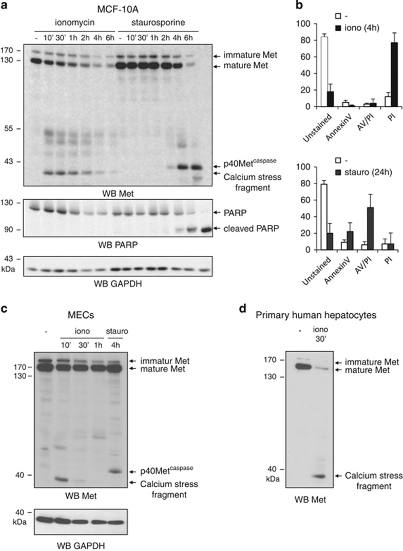
Figure 1.
Ionomycin induces necrotic cell death and generation of a 40-kDa Met fragment. (a) MCF-10A cells were grown for 24 h, serum starved overnight, and treated with 1 μM ionomycin (iono) or 1 μM staurosporine (stauro) for the indicated time. (b) MCF-10 A cells were treated with 1 μM ionomycin or 1 μM staurosporine then stained with Annexin V-fluorescein isothiocyanate (FITC) and PI. Percentages of unstained cells and of cells stained with Annexin V, PI, or both (AV/PI) are shown. Mouse mammary epithelial cells (MECs) (c) or human primary hepatocytes (d) were treated with 1 μM ionomycin or 1 μM staurosporine for the indicated time. (a, c and d) Cell lysates were analyzed by western blotting with an antibody directed against the kinase domain of human Met, against PARP to assess caspase activation, or against GAPDH to assess loading. Arrows indicate full-length Met, p40Metcaspase, and the calcium-stress-induced fragment

