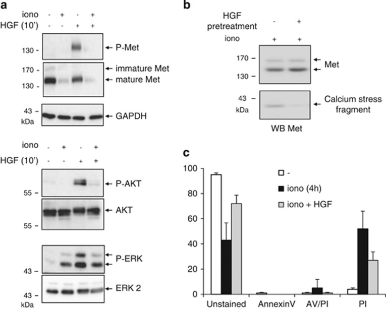
Figure 2.
Calcium-stress-induced Met degradation impairs Met survival signaling. (a) MCF-10 A cells were grown for 24 h, serum starved overnight, treated for 1 h with 1 μM ionomycin (iono), and then stimulated or not 10 min with 10 ng/ml HGF/SF. (b and c) MCF-10 A cells were grown for 24 h, serum starved overnight in the presence of vehicle or 10 ng/ml HGF/SF, then treated for 1 h with 1 μM ionomycin and (b) analyzed by western blotting or (c) stained with Annexin V-fluorescein isothiocyanate (FITC) (AV) and PI. (a and b) Cell lysates were analyzed by western blotting with antibodies directed against the kinase domain of human Met, AKT, ERK, or the phosphorylated form thereof (P-Met, P-AKT, or P-ERK) and against GAPDH to assess loading. Arrows indicate the positions of the respective detected proteins

