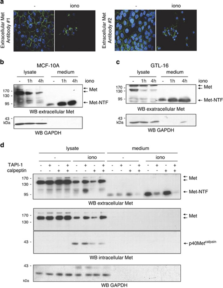
Figure 5.
Ionomycin (iono) treatment increases Met shedding. (a) MCF-10A cells were grown on glass coverslips, serum starved overnight, and treated with vehicle or 1 μM ionomycin for 4 h. Immunofluorescence staining was performed with two different antibodies directed against the Met extracellular region and the nuclei were stained with Hoechst. (b) MCF-10 A and (c) GTL-16 cells were grown for 24 h, serum starved overnight, and treated with 1 μM ionomycin for the indicated time. Both cell lysates and an equal volume of conditioned medium were analyzed by western blotting with an antibody against the Met extracellular region. (d) MCF-10A and GTL-16 cells were grown for 24 h, serum starved, and pretreated overnight with TAPI-1 and/or calpeptin, and treated for 1 h with 1 μM ionomycin. Cell lysates and conditioned medium were analyzed by western blotting with an antibody against the Met extracellular region, the Met kinase domain (intracellular Met), and GAPDH to assess loading. Arrows indicate positions of full-length Met, Met-NTF, and p40Metcalpain

