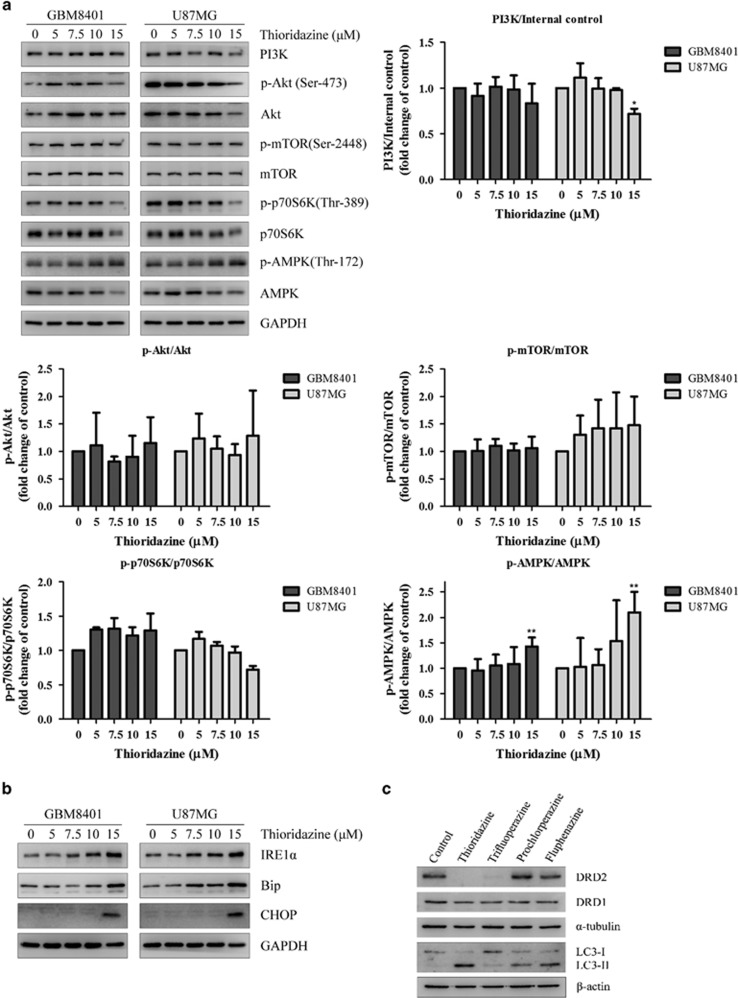
Figure 3.
Thioridazine modulates PI3K/Akt/p70S6K signaling pathways and induces ER stress in GBM cells. (a) GBM8401 and U87MG cells were treated with thioridazine at 5, 7.5, 10 and 15 μM for 24 h. PI3K, phospho-Akt (Ser-473), phospho-mTOR (Ser-2448) and phospho-S6K (Ser-424) and phospho-AMPK (Thr-172) were detected by western blotting. GAPDH was used as an internal control. The quantification of the western blotting intensity was normalized to internal control intensity using ImageJ. Bar graph represents the mean of triplicates±S.D. *P<0.05, **P<0.01 compared with the control group. (b) GBM8401 and U87MG cells were treated with thioridazine at 5, 7.5, 10 and 15 μM for 24 h. ER stress markers IRE1α, Bip and CHOP were detected by western blotting. GAPDH was used as an internal control. (c) GBM8401 cells were treated with different antipsychotic drugs at 10 μM for 24 h. DRD2, DRD1, LC3-I and LC3-II were detected by western blotting. α-tubulin and β-actin were used as internal controls

