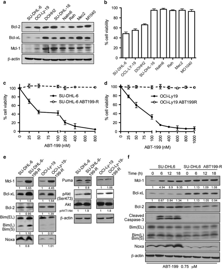
Figure 1.
DLBCL cells with low MCL-1 and/or BCL-xL expression are sensitive to ABT-199 and develop resistance after chronic ABT-199 exposure by upregulating MCL-1, BCL-xL, and p-AKT levels. (a) Expression levels of the pro-survival proteins MCL-1, BCL-2, and BCL-xL in DLBCL (SU-DHL-6, OCL-LY-19, SU-DHL-16), follicular lymphoma (DOHH2), acute lymphoblastic leukemia (Nalm6, Reh), and CLL (Mec2, MO1040) cell lines. β-actin was used as the loading control. (b) The indicated cell lines were treated with 50 nM ABT-199 for 48 h. The viability shown represents the percentage of live cells relative to control cells treated with dimethyl sulfoxide. Parental and ABT-199-R (c) SU-DHL-6 and (d) OCl-Ly-19 cells were treated with the indicated concentrations of ABT-199 for 48 h and cell viability was determined by Annexin V-PI staining. Control cells were treated with dimethyl sulfoxide. (e) Expression levels of: (i) pro-survival proteins MCL-1, BCL-2, and BCL-xL, (ii) pro-apoptotic proteins BIM, NOXA, and PUMA, and (iii) total AKT and p-AKT (Ser473) in untreated parental and ABT199-R-derivative DLBCL cell lines at the indicated time. SU-DHL-6 parental and resistant cells. Both panels represent one experiment with BCL-2 and β-actin serving as loading controls. (f) Cells were treated with ABT-199 for the indicated time. MCL-1, BCL-2, BCL-xL, BIM, NOXA, PUMA, and cleaved caspase-3 were determined by immunoblotting. β-actin was used as a loading control. Standard deviation (S.D.) is indicated in b–d as error bars (N=3). The experiments in a, e, and f are representative of three independent experiments. Numbers below blots indicate increase in protein levels as determined by ImageJ quantification, which in case of pAKT was normalized to total AKT levels

