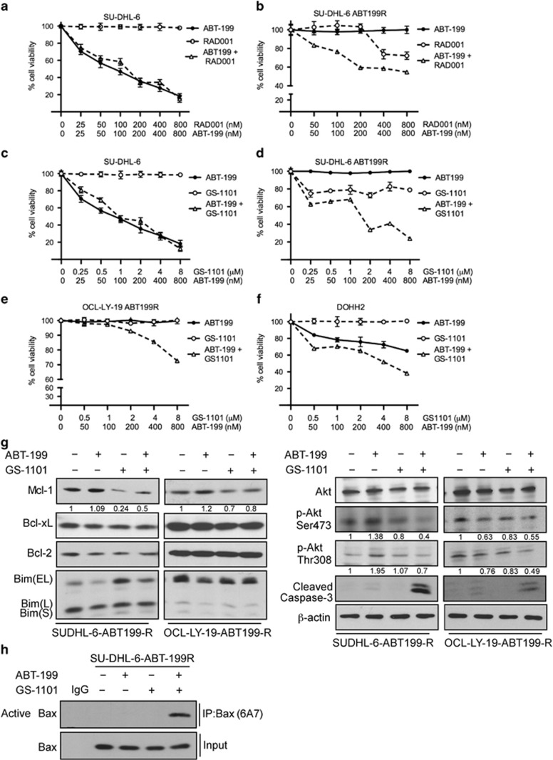
Figure 6.
GS-1101 in combination with ABT-199 targets the PI3K pathway, sensitizing ABT-199-R cells by MCL-1 downregulation and BAX activation. Parental and ABT-199-R-derivative DLBCL cell lines SU-DHL-6 (a–d), OCL-LY-19 ABT199-R (e) and the FL cell line DOHH2 (f) were treated for 24 h with the indicated concentration of ABT-199, RAD001, and GS-1101, alone or in combination. Cell viability was determined by staining with Annexin V-PI, and represented as the percentage relative to control cells treated with dimethyl sulfoxide. Standard deviation (S.D.) is indicated in a–e by error bars (N=3). (g) Expression levels of MCL-1, BCL-xL, BCL-2, BIM, p-AKT (Ser473), p-AKT (Thr308), AKT, and cleaved caspase-3 in SU-DHL-6 ABT199-R cells treated with ABT-199 (400 nM) and GS-1101 (4 μM) alone or in combination, and OCL-LY-19 ABT199-R treated with ABT-199 (400 nM) and GS-1101 (8 μM), alone or in combination, for 24 h. β-actin was used as the loading control. (h) ABT-199-R SU-DHL-6 cells treated with ABT-199R (400 nM), GS-1101 (4 μM), alone or in combination, were lysed with 1% CHAPS buffer, and active BAX was immunoprecipated with BAX 6A7 and detected using BAX N20 antibodies following immunoblotting. The experiments in g and h are representative of three independent experiments

