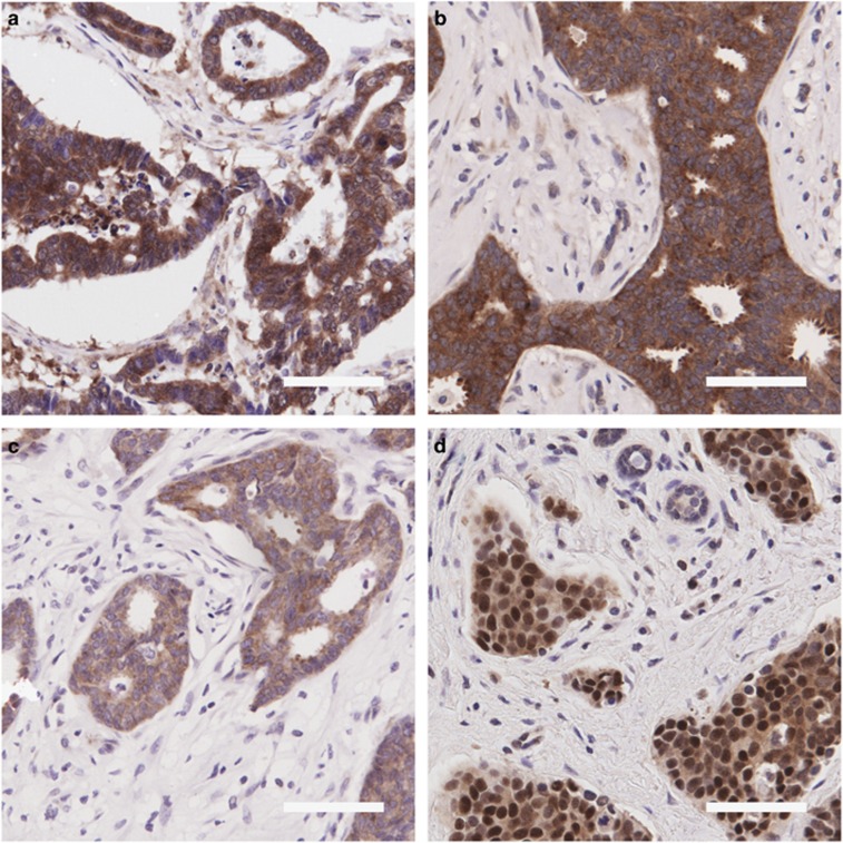
Figure 1.
Representative immunohistochemistry images. Photomicrographs of representative TMA cores were taken. (a) eIF4E (strong cytoplasmic staining), (b) eIF4A1 (strong cytoplasmic staining), (c) eIF4B (moderate cytoplasmic staining) and (d) PDCD4 (strong nuclear staining and moderate cytoplasmic staining). Scale bar=100 μm

