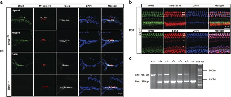
Figure 1.
Bmi1 expressed in auditory hair cells and supporting cells. (a) Immunofluorescence staining showed Bmi1 expression in the apical, middle and basal turns in the Corti's organ of neonatal (P0) WT mice. Myosin 7a and Sox2 were used as hair cell and supporting cell markers, respectively. (b) Bmi1 expressed in the cochlear epithelium of P30 WT mice. (c) Typical PCR data of genotyping. Scale bars: 40 μm (a); 20 μm (b). OHC, outer hair cell; IHC, inner hair cell; DC, Deiters' cell; IP, inner pillar cell; OP, outer pillar cell

