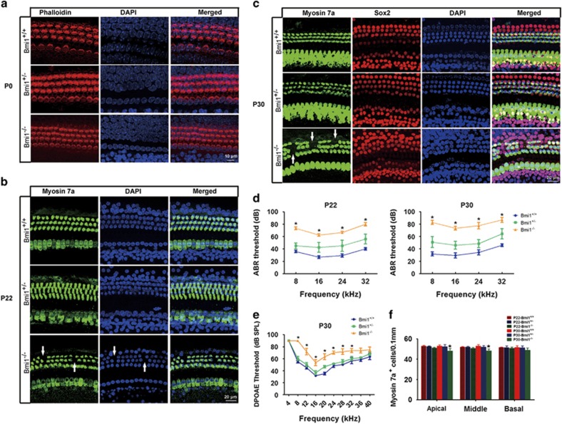
Figure 2.
Bmi1−/− mice showed severe hearing loss and patched auditory hair cell loss. (a) Phalloidin staining showed that the number and morphology of cochlear hair cells are similar among neonatal Bmi1+/+, Bmi1+/− and Bmi1−/− mice. (b) Myosin 7a staining showed dot loss of outer hair cells in P22 Bmi1−/− mice. (c) Myosin 7a and Sox2 immunofluorescence staining showed patched loss of outer hair cells in P30 Bmi1−/− mice. (d) ABR measurement revealed that hearing threshold at 8, 16, 24 and 32 KHz significantly increased in Bmi1−/− mice compared with WT littermate at P22 and P30. (e) DPOAE measurement showed the function of outer hair cells was impaired in P30 Bmi1−/− mice. (f) Statistical data of myosin 7a positive cells in the cochlea of P22 and P30 mice. Scale bars: 10 μm (a); 20 μm (b and c). *P<0.05 versus Bmi1+/+ group. n=6 for each group

