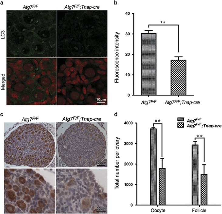
Figure 3.
Oocytes and follicles over-loss in 3-day-old Atg7 knockout mice ovaries. (a) Autophagy was severely disrupted in 3-day-old Atg7F/F;Tnap-cre mice (n=3) ovaries by immunostaining of LC3. LC3 (in green), nucleus (in red). (b) Quantification of LC3 fluorescent intensity in panel (a). (c) Morphological analysis of 3-day-old ovaries after Atg7 knockout. Germ cell was indicated by immunohistochemical detection of MVH. (d) Quantification of oocyte and follicle numbers in 3-day-old mice ovaries of Atg7F/F;Tnap-cre mice (n=3) and the control (n=3). Oocytes at least surrounded by three cells were counted as follicles. Data are presented as the mean±S.E.M. **P<0.01

