Figure 5.
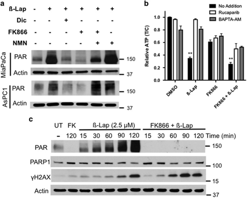
FK866 addition abrogated PAR formation and increased DSBs in β-lap-treated MiaPaca2 cells. (a) NQO1+ MiaPaca2 or AsPC1 PDA cells were pretreated with FK866 (8 nM, 24 h), then β-lap (2.5 μM) was added in the presence or absence of dicoumarol (Dic, 50 μM) or NMN (500 μM) for 20 min. Cell extracts were analyzed for PAR formation. Dicoumarol inhibits NQO1 activity thereby preventing PARP1 hyperactivation. β-Lap-induced PAR formation was abrogated by FK866 (8 nM) and rescued with NMN (500 μM). Actin levels were monitored as loading controls. (b) MiaPaca2 cells were pretreated with or without FK866 (8 nM) and then exposed to β-lap (3 μM) in the presence or absence of (i) a PARP1 inhibitor (Rucaparib (AG014699), 20 μM) or (ii) a calcium chelator (BAPTA-AM) for 2 h; Rucaparib or BATPA-AM treatments prevent PARP1 hyperactivation in β-lap-treated cells.16, 29, 42 ATP levels were measured after the 2 h co-treatment. Student's t-tests were performed comparing β-lap or FK866+β-lap versus addition of Rucaparib (AG014699) (n=3 ± S.D.). **P <0.01. (c) MiaPaca2 cells were pretreated or not with FK866 (8 nM, 24 h), then exposed to β-lap (2.5 μM) and PAR and γ-H2AX formation were monitored at various times during the 2 h exposure by western blot. Mean γH2AX band intensities, as a measure of DNA double-strand break formation, were normalized to actin and levels graphed over time (mins) in Supplementary Figure S1D. Reduced PARP1 activity, as a result of lowered NAD+ levels from FK866 exposure, resulted in more rapid induction of γ-H2AX

