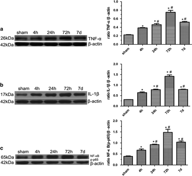
Figure 4.
Expression of TNF-α, IL-1β and NF-κB in the brains of MCAO rats. Western blot confirmed that protein levels of TNF-α, IL-1β and phospho-NF-κB p65(NF-κB p-p65) increased as early as 4 h after reperfusion, further increased at 24 h, peaked at 72 h and decreased at 7 days, respectively (*P<0.001, compared with the controls; #P<0.001 compared with the previous time point) (a–c). Five rats in each group were used for western blot (n=5), sham, sham-operated control rats

