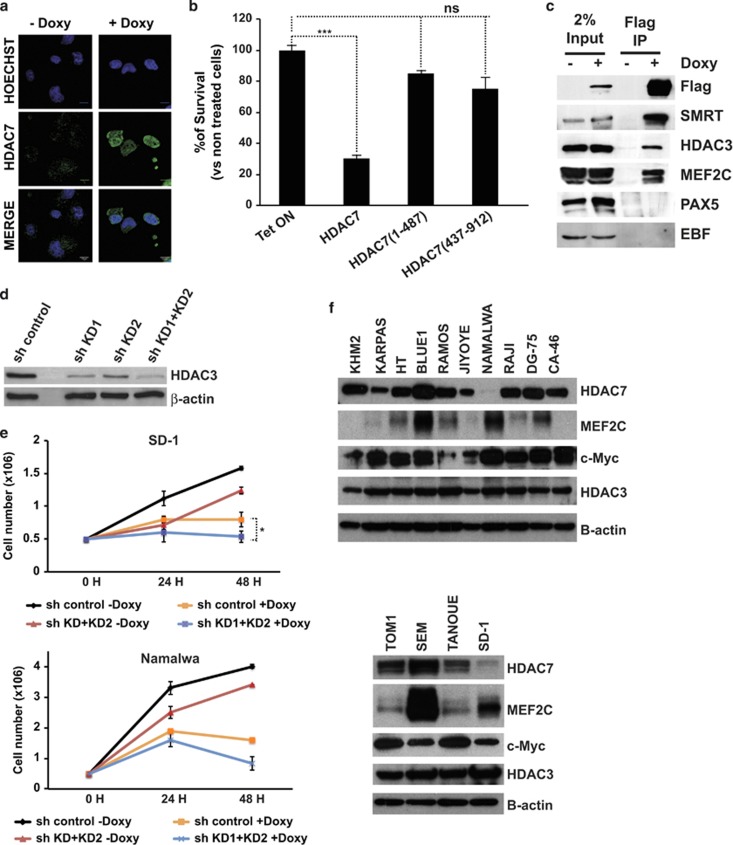
Figure 6.
HDAC7 interacts with MEF2C, HDAC3 and SMRT and is localized in the nucleus. (a) Namalwa-Tet-On-Tight-HDAC7 cells were treated, or not, with doxycycline for 24 h. The subcellular localization of HDAC7 was determined by immunofluorescence. (b) Mean±S.E.M. of the percentage of survival cells from three independent MTT assays performed in triplicate of Namalwa cells expressing empty vector, wild-type HDAC7, and HDAC7 (1–487) and HDAC7 (438–915) deleted forms treated, or not, with doxycycline. ***P<0.001. (c) Cell lysates from Namalwa-Tet-On-Tight-HDAC7 treated, or not, with doxycycline were immunoprecipitated with anti-M2 agarose beads and analyzed by western blotting with the indicated antibodies. (d) Namalwa-Tet-On-Tight-HDAC7 cells were transduced with lentiviral vectors for the expression of the control shRNA or the shRNAs targeting HDAC3, and GFP-positive cells were purified by flow cytometry. HDAC3 protein levels were assessed by western blot. (e) SD-1-Tet-On-Tight-HDAC7 and Namalwa-Tet-On-Tight-HDAC7 cells transduced with either pLKO.1-shRNA control or pLKO.1-shHDAC3KD1+KD2 lentiviral vectors were cultured and treated, or not, with doxycycline. At the indicated times after treatment, the cell number was assessed by cell counting. Trypan blue-dyed cells were omitted from the cell counts. Means±S.D. of the three independent experiments performed in triplicate. (f) Western blots for HDAC7, MEF2C, c-Myc and HDAC3 in B-ALL and lymphoma cell lines

