Figure 1.
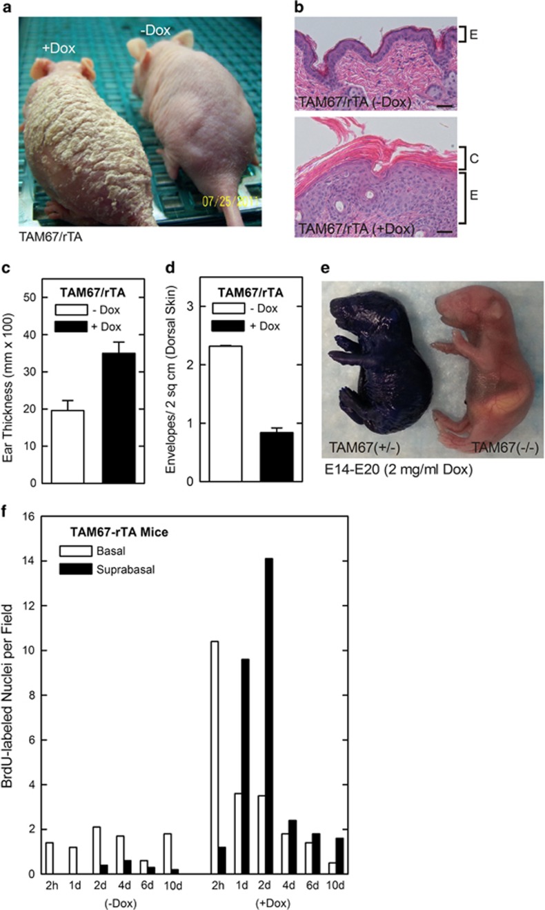
Impact of suprabasal epidermal AP1 factor inactivation on epidermal phenotype . (a and b) TAM67-rTA mice were treated with 0 or 2 mg/ml doxycycline for 7 days and the mice were photographed and skin sections were processed and stained with hematoxylin and eosin. E indicates the epidermis and C the cornified layers. (c) Increased ear thickness in TAM67-FLAG-positive epidermis. Ear thickness was monitored using calipers at 7 days after initiation of doxycycline treatment. (d) Reduced cornified envelope number in TAM67–FLAG-positive epidermis. Epidermis scale was collected from TAM67-rTA mice treated as above. The scale was then boiled in detergent and reducing agent and surviving envelope structures were counted. The values are mean±S.E.M. of three separate experiments. Envelope number is significantly reduced (P<0.001, n=3). (e) TAM67 expression compromises barrier function. Pregnant female mice were treated with 2 mg/ml doxycycline beginning on E14 and the embryos were removed and stained with toluidine blue at E20. All TAM67-positive mice displayed compromised barrier function. (f) TAM67 expression is associated with increased cell proliferation. Adult TAM67-rTA mice were treated with 0 or 2 mg/ml doxycycline and on day 4 were injected IP with 50 mg of BrdU per kg body weight. At 2 h to 10 days animals were killed and the epidermis was sectioned and stained with anti-BrdU. The number of basal and suprabasal BrdU-positive cells were counted at each time point. Similar findings were observed in each of two experiments

