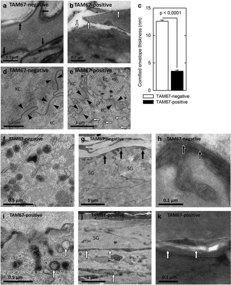
Figure 2.
Attenuated cornified envelope, keratin filament and desmosome formation and altered lipid secretion in TAM67-positive epidermis. (a and b) Electron microscopy reveals uniform cornified envelopes (black arrows) in stratum corneum of TAM67-negative as compared with less-developed envelopes (white arrows) in TAM67-positive epidermis. (c) Quantification of the cornified envelope thickness. The values are mean+S.E.M. (d and e) TAM67-negative epidermis demonstrates compact desmosomes flanked with keratin filaments (black arrowheads) compared with disorganized desmosomes (black arrowheads) and haphazardly arranged keratin filaments (white arrows) in TAM67-positive epidermis. KC indicates keratinocyte. Epidermis from TAM67-negative mice is characterized by lamellar bodies of uniform size filled with stacked membranous lipid contents (f), lipid secretion at the SG–SC junction (g, black arrows) and processing of secreted lipid into extended membrane arrays (h, ruthenium stained, black arrows). In contrast, TAM67-rTA epidermis displays abnormal lamellar body contents (i, white arrows), premature lipid secretion in the middle SG layers (j, white arrows) and incomplete post-secretory processing of secreted lipid (k, white arrows). SG, stratum granulosum; SC, stratum corneum

