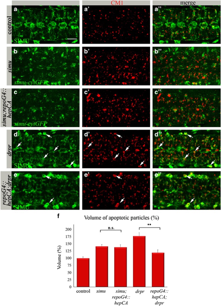
Figure 6.
dJNK signaling in glia does not rescue the simu phenotype, whereas it substantially rescues the drpr phenotype. (a–e″) Projections from confocal stacks of the CNS at embryonic stage 16, ventral view. Apoptotic cells in red (CM1), whereas glia (in green) are labeled with different markers. Bar 20 μm. In control repoGal4 embryo (a–a″) apoptotic particles are mostly inside anti-SIMU-labeled glia. In the simu mutant (b–b″), many apoptotic particles are outside glia labeled with simu-cytGFP. (d–d″) In the drpr mutant, apoptotic particles accumulate inside glia and macrophages (arrows). (c–c″ and e–e″) Rescue experiments of simu (c–c″) or drpr (e–e″) mutants by dJNK signaling in glia (repoGal4::hepCA). No rescue is detected in the simu mutant (c–c″ and f) but substantial rescue is detected in the drpr mutant (e–e″ and f). Many apoptotic particles accumulate in macrophages outside the CNS (arrows) but not in glia (e″). (f) Quantification of phenotypic rescue of simu and drpr mutants by repoGal4::hepCA. Columns represent mean total volume of apoptotic particles within confocal stacks of the CNS, ±S.E.M., n=5–9; asterisks indicate statistical significance versus simu and drpr mutants, as determined by the Student's t-test, **P<0.002, NS (not significant) P>0.05

