Figure 2.
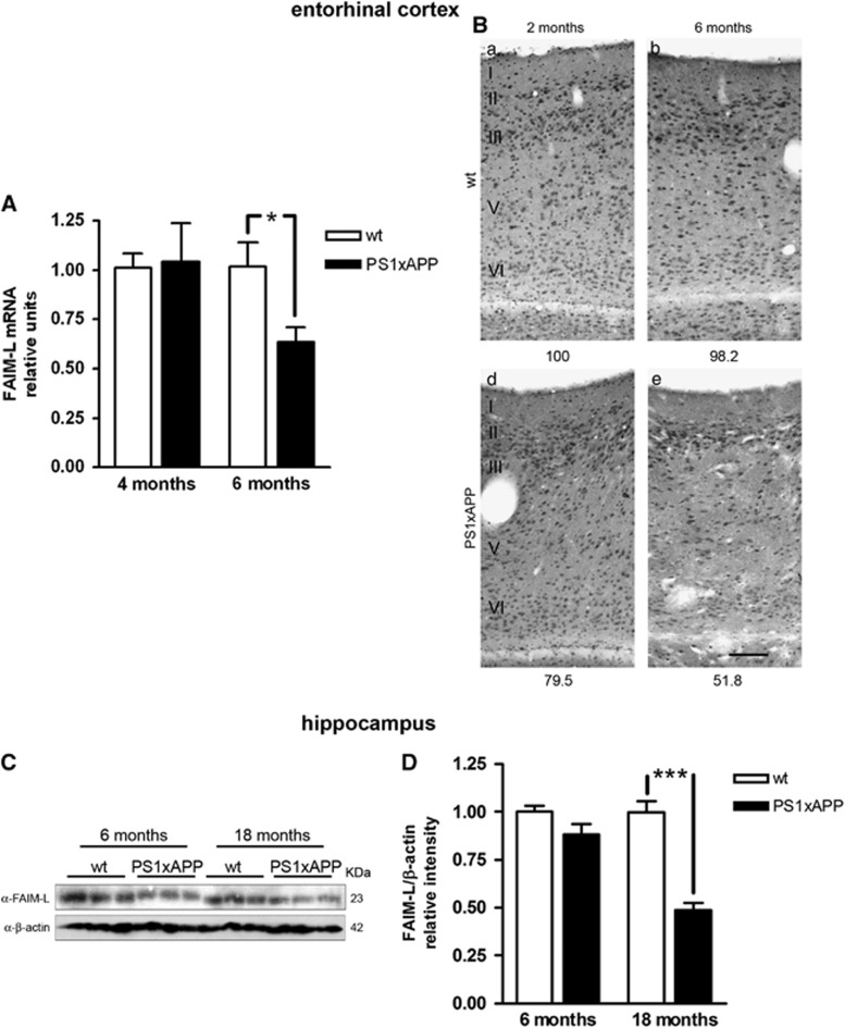
Reduction of FAIM-L expression in transgenic PS1xAPP animals. (A) FAIM-L mRNA levels in laser-microdissected entorhinal cortex. *T-test with P=0.039. Data are mean±S.D. of three independent experiments. (B) Immunodetection of FAIM-L expression in the entorhinal cortex. In the entorhinal cortex, WT animals display a similar immunostaining pattern for FAIM-L antibody at the ages analyzed (2 and 6 months old), without clear differences between superficial (I–III) and deep (IV–VI) layers. The PS1xAPP transgenic animals, however, present a marked reduction in FAIM-L labeling, slightly at 2 months of age, but patent at 6 months. The decline in FAIM-L immunoreactivity does not follow any apparent pattern, showing a reduction in all the layers, possibly less pronounced in neurons in layers II–III. Numbers at the bottom of the micrographs indicate the relative integrated density of the FAIM-L labeling. Scale bar,100 μm. (C) Western blot of FAIM-L in samples from WT and transgenic animals at different ages. Three different animals were used to obtain homogenates samples for each condition analyzed. (D) Protein FAIM-L quantification in hippocampus using β-actin as a loading control. Data are mean±S.D. of three independent western blots with three animal samples for condition. T-test ***P<0.0001

