Figure 1.
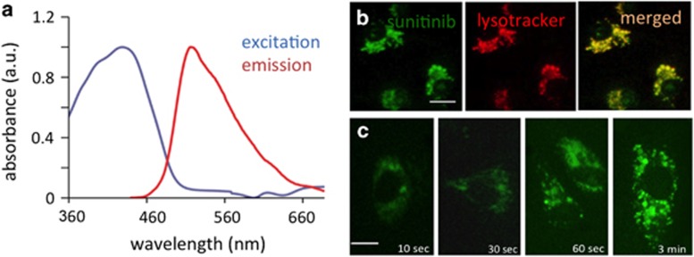
Spectral properties of sunitinib and lysosomal accumulation in endothelial cells. (a) Normalized absorption and emission spectra of sunitinib in 0.1% DMSO in 0.9% NaCl. (b) Colocalization of sunitinib (green) and lysotracker (red) in cultured human umbilical vein endothelial cells (HUVEC). Bar in left panel represents 10 μm. (c) Rapid uptake and lysosomal accumulation of sunitinib in HUVEC. Bar in left panel represents 5 μm

