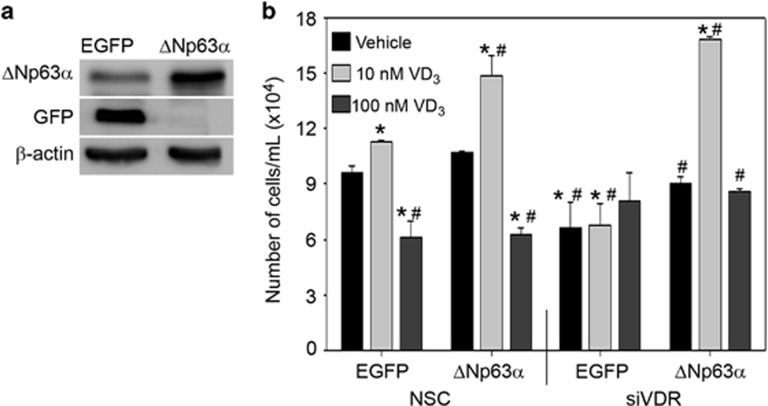
Figure 6.
ΔNp63α rescues reduction in VD3-mediated cell proliferation following loss of VDR. (a) The expression of eGFP and ΔNp63α were confirmed in HaCaT-eGFP and HaCaT-ΔNp63α stable cells via immunoblot analysis. (b) HaCaT-eGFP and HaCaT-ΔNp63α stable pools were transfected with non-silencing control (NSC) or siVDR followed by treatment with vehicle control, 10 nM or 100 nM VD3 for 24 h. Cell viability was measured following VD3 treatment by trypan blue cell exclusion. Error bars represent standard deviation from the mean. *P values≤0.05 compared with vehicle control EGFP expressing HaCaT cells. #P values≤0.05 compared with 10 nM EGFP expressing HaCaT cells

