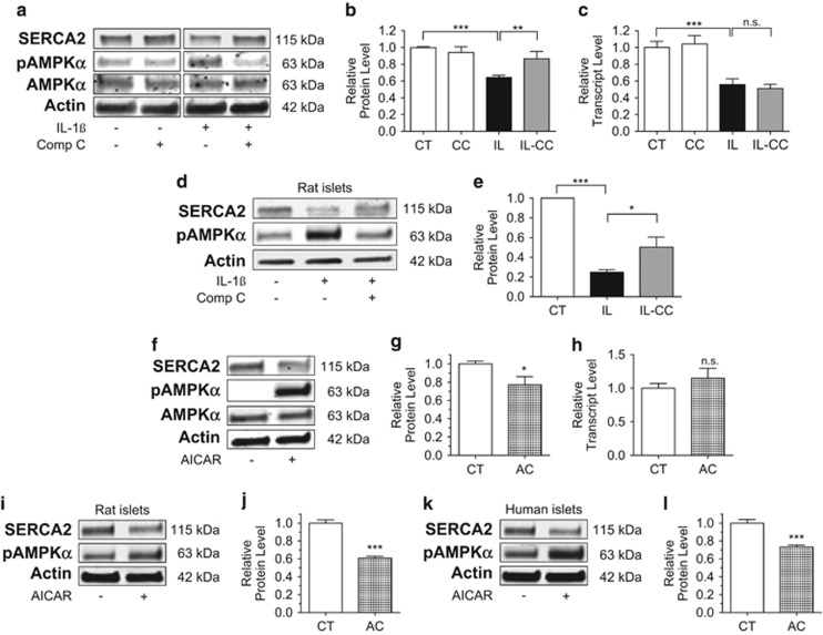
Figure 3.
Activation of AMPKα Th173 leads to a loss of SERCA2 protein expression. INS-1 cells (a–c) or isolated rat islets (d–e) were treated with dimethyl sulfoxide (DMSO) (CT) or 5 ng/ml IL-1β (IL) combined with or without 10 μM of the AMPK inhibitor CC for 24 h. Total protein was isolated, and immunoblot was performed using antibodies against SERCA2, phosphorylated AMPKα Th173 (pAMPKα), total AMPK and actin. Quantitative SERCA2 protein levels are shown graphically (b and e). Total mRNA was isolated from INS-1 cells treated with CT, IL and IL-CC, and reverse-transcribed RNA was subjected to quantitative real-time PCR (qRT-PCR) for quantification of SERCA2b and actin transcript levels (c). Next, INS-1 cells or isolated rat islets and human islets were treated with and without the AMPK activator AICAR (AC) at 2 mM for 24 h (f–l). Total protein and mRNA were isolated; immunoblot was performed using antibodies against SERCA2, pAMPKα, AMPK and actin. Quantitative protein levels of SERCA2 are shown graphically (g, j and l). Reverse-transcribed RNA was subjected to qRT-PCR for quantification of SERCA2b and actin transcript levels (h). Indicated comparisons are significantly different (*P<0.05, **P<0.01 and ***P<0.001), or results are statistically different from control conditions (g, j and l)

