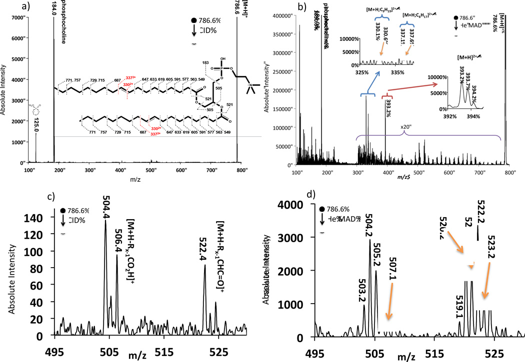
Figure 1.
Comparison spectra of a) even electron CID of [9E-DOPC+H]1+ (18:1/18:1) and b) odd-electron He-MAD of the same 1+ protonated lipid. c) and d) show expanded regions of the loss of sn-1 and sn-2 acyl chains for CID and He MAD, respectively. The inset in a) also shows possible cleavages and theoretical masses for fragmentation with no H-atom transfers. The masses in the insert may not agree with those shown in the spectra due to hydride shifts or rearrangements.

