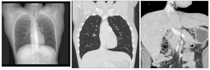
Fig. 1.
(Left) An x-ray projection image of the chest. (Middle) a CT slice image of the same patient, cutting vertically through the heart and lungs; the gray scale window is [−1200 HU, 400 HU]. (Right) a zoom and pan of the same CT slice image showing the patient’s lower abdomen [−150 HU, 200 HU]. The gray scale units are in the Hounsfield (HU) scale, where −1000 HU and 0 HU correspond to air and water, respectively. The CT lung image, displayed in a wide gray scale window, faithfully represents the structures in this slice without interference from overlapping lung tissue or ribs, seen in the x-ray projection. Such a representation is needed to view subtle lesions in the lung. Note that the image of the heart is sharp—an indication of the high temporal resolution of diagnostic CT. The CT abdomen image, displayed in a narrow gray scale window, shows the ability of CT to resolve low-contrast structures with high spatial resolution. Images courtesy of Alexandra Cunliffe, Ph.D.

