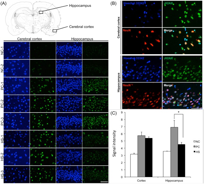
Fig 9. In vivo validation of hippocampal avoidance with radiation in rats treated with brain irradiation with hippocampal avoidance.
A. Representative immunofluorescence images of rats treated with no irradiation (Control), whole brain irradiation (WBRT), and hippocampal avoidance brain irradiation (HA). For each rat, a section of brain from the cortex (not spared) and a section from the hippocampus (granule cell layer of the dentate gyrus) is shown. A brain map (top) indicates the locations where the images of the cortex and hippocampus are taken. Scale bar = 250 microns. B. Anti-phospho-Histone H2A.X antibody immunofluorescence signal intensity analysis. The radiation-induced phosphorylation of Histone H2A.X in the hippocampus is significantly decreased when the brain is irradiated with hippocampal avoidance, * P = 0.012 (by student t-test). C. Immunofluorescence images of the cortex and hippocampus of a rat treated with WBRT. Red = NeuN (a neuron-specific marker), Green = phosphorylation of Histone H2A.X. Scale bar = 125 microns.

