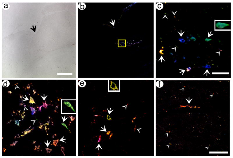
Fig. 4.
pVGLUT1brainbow supports labeling of POR cortex neurons and axons, and distant axons in PER cortex, with different hues. Rats were sacrificed at 4 days (A–E) or 8 days (F) after gene transfer, brains were sectioned, and confocal stacks were analyzed. (A) A brightfield, low power view of POR cortex in a section proximal to the injection site. The arrow indicated the rhinal sulcus. (B) A low power view of Brainbow labeled neurons in the same section as in (A). (C) A high power view of the boxed area from (B). Neurons and proximal axons that contain different hues are shown. Arrows, cell bodies; arrowheads, axons. (D and E) High power views of neurons and proximal axons, in POR cortex, that contain different hues, from different sections. Some of the labeled cell bodies contain proximal processes, particularly in (D). Also, some axons that are not connected to a cell body are visible. (F) A high power view of PER cortex that shows one axon for ~50 um in length (arrow) and other axons in cross section (arrowheads). Scale bars: (A and B) 250 μm, (C–E) 50 μm, (F) 50 μm.

