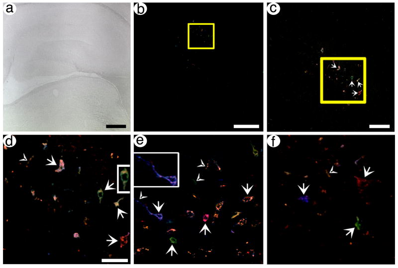
Fig. 5.
pVGLUT1brainbow can label neurons and proximal axons in the hippocampal dentate gyrus with different hues. Rats were sacrificed at 4 days after gene transfer, and confocal stacks were collected. (A) A brightfield, low power view of the hippocampus and adjacent areas in a section proximal to the injection site. (B) A low power view of Brainbow labeled neurons in the same section as in (A). (C) A medium power view of the boxed area from (B) showing Brainbow labeled cells. Arrows, cell bodies. (D) A high power view of the boxed area from (C) showing Brainbow labeled neurons and proximal axons. Arrows, cell bodies; arrowheads, axons. (E and F) High power views of Brainbow labeled neurons and proximal axons from different sections that contain the dentate gyrus. Some of the labeled cell bodies contain proximal processes, particularly a blue neuron on the left side of (E) contains an axon that extends for ~50 μm. Also, some axons that are not connected to a cell body are visible. Scale bars: (A) 500 μm, (B) 250 μm, (C) 100 μm, (D–F) 50 μm.

