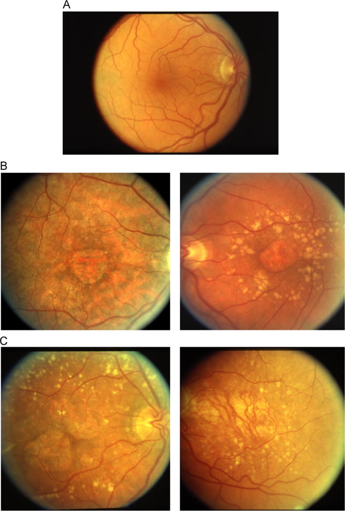
Fig. 1.

Row A shows a color fundus image of a healthy eye, with an absence of AMD and geographic atrophy. Row B shows two examples of GA that were labeled as unambiguous by our retina specialist. Row C shows two examples of GA that were labeled as ambiguous by our retina specialist. (For interpretation of the references to color in this figure caption, the reader is referred to the web version of this paper.)
