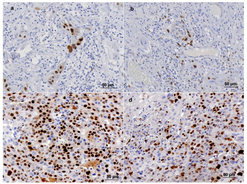
Figure 1.
Cyclin D1 and Ki-67 immunohistochemical staining in tongue squamous cell carcinoma. a and b - Cyclin D1 staining (≤ 19 stained cells per field) and Ki-67 staining (≤ 28 stained cells per field) in the same tumor, respectively; c and d - Cyclin D1 staining (> 19 stained cells per field) and Ki-67 staining (> 28 stained cells per field) in the same tumor respectively. 400X magnification.

