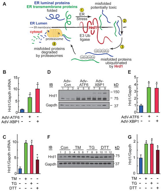
Figure 1. Characterization of Hrd1 Expression in Cardiac Myocytes.
A, Diagram of ER-associated protein degradation (ERAD). B, D and E, Cultured cardiac myocytes were treated with AdV-Con, AdV-ATF6, or AdV-XBP1 for 48h. Hrd1 and Gapdh mRNA B, or protein D, were measured by qRT-PCR or immunoblotting, respectively. E, Densitometry of the immunoblot shown in D. C, F and G, Cultured cardiac myocytes were treated with tunicamycin (TM) 10 μg/ml, thapsigargin (TG) 1 μM, or dithiothrietol (DTT) 1mM for 20h. Hrd1 and Gapdh mRNA C, or protein F, were measured by qRT-PCR or immunoblotting, respectively. G, Densitometry of the immunoblot shown in F. * = p ≤ 0.05 different from control.

