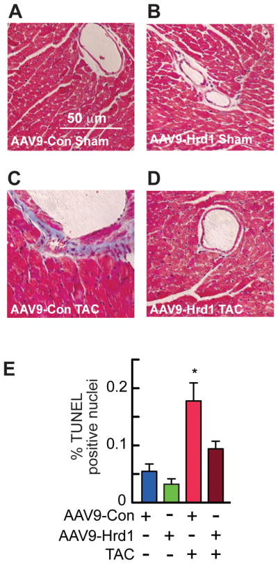
Figure 7. Histology and TUNEL Analyses of Mouse Hearts Treated with AAV9-Con and AAV9-Hrd1.

A–D, Sections of hearts from mice treated with either AAV9-Con or AAV9-Hrd1, and then subjected to sham or TAC surgery, as described in the Experimental Protocol shown in Fig. 6A, were stained with Masson’s trichrome to examine fibrosis (blue). E, Sections of hearts from mice treated with either AAV9- Con or AAV9-Hrd1, and then subjected to TAC were analyzed for apoptosis by TUNEL staining, then quantified to determine % of nuclei that were TUNEL-positive. * = p ≤ 0.05 different from all other values.
