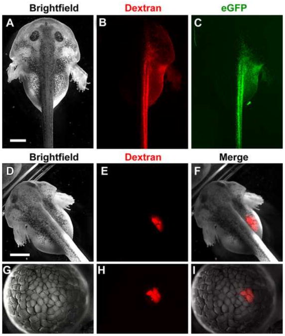
Figure 6.
L. laevis embryos are amenable to microinjection of exogenous reagents for fate-mapping and expression of synthetic mRNA. Embryos were injected at the 2-cell stage (A–C), 8-cell stage (D–F) or 128-cell stage (G–I) with red fluorescent dextran and/or capped eGFP mRNA. By 48 hours post fertilization (hpf), injection into a single blastomere at the 2-cell stage results in predominantly unilateral labeling (B) and expression of eGFP (C, green), while injection into a single blastomere at the 8-cell stage results in fluorescence limited to the developing gut (E). Injection of dextran into a single blastomere at the 128-cell stage labels a limited region of only 4 cells by the mid-blastula stage (H). Merge of brightfield and fluorescent images in (F, I). Scale bars = 1 mm.

