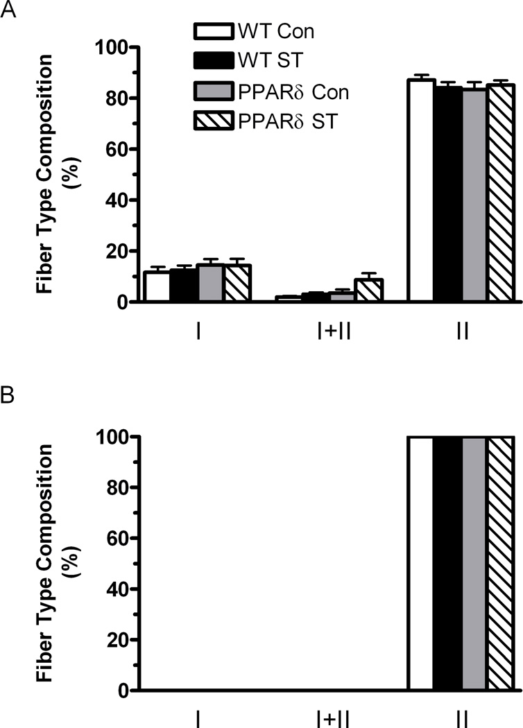
Figure 5.
Fiber type composition determined by MHC immunohistochemistry. The data are expressed as a percentage of the total number of fibers sampled in the deep (A) (close to the bone) and superficial (B) (away from the bone) regions of the MG from WT-Con, WT-ST, PPARδ-Con, and PPARδ-ST groups. Values are means ± SEM.

