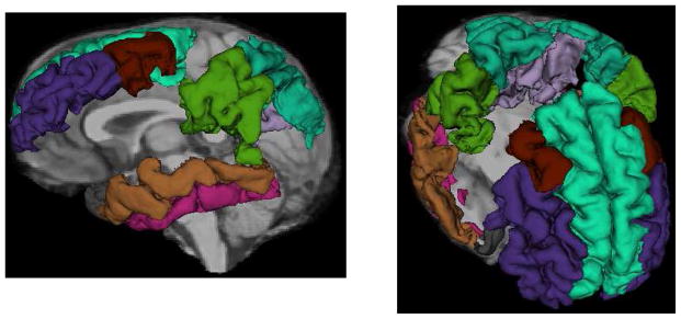
Figure 1.
FreeSurfer regions-of-interest representing the 8 regions comprising the AD cortical signature superimposed on a single T1-weighted MRI scan. Labeled regions include the medial temporal gyrus (brown), temporal pole (grey), inferior temporal gyrus (pink), supramarginal gyrus (dark green), superior parietal lobule (teal), precuneus (lavender), middle frontal gyrus (purple: rostral, rust: caudal), and superior frontal gyrus (light teal)

