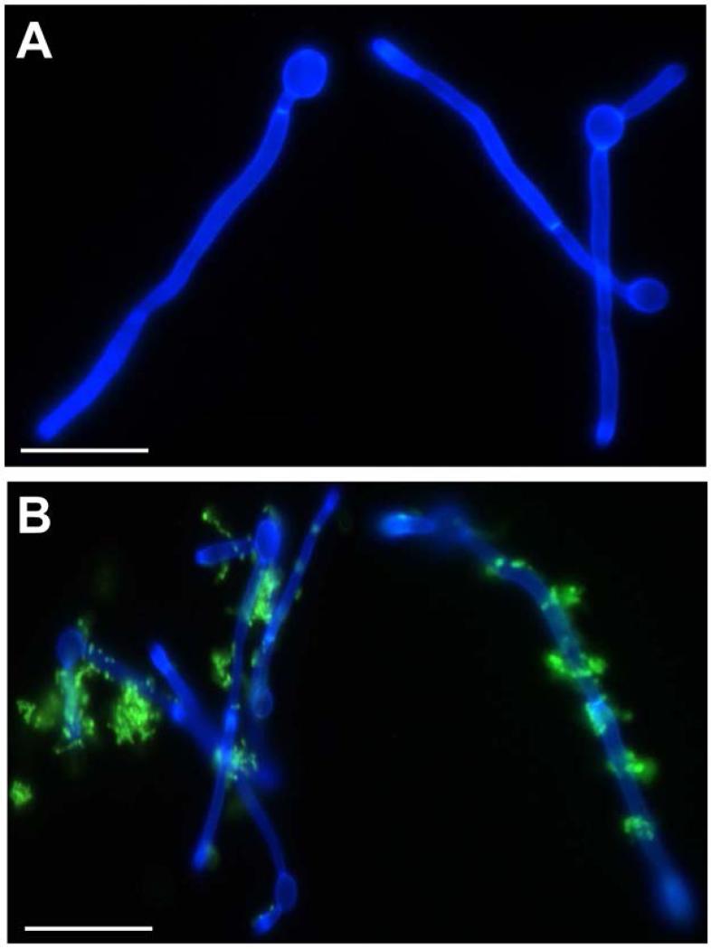
Fig. 1.
Light micrograph images of C. albicans SC5314 cells interacting with S. gordonii DL1 cells. S. gordonii cells were fluorescently labelled with fluorescein isothiocyanate (FITC) and incubated with filamentation-induced (2 h at 37°C) C. albicans for 1 h at 37°C with gentle agitation. Calcofluor white (0.3 μg ml−1) was added to fluorescently label C. albicans. Panel A, C. albicans alone; Panel B, C. albicans and S. gordonii (green). Scale bar 5 μm.

