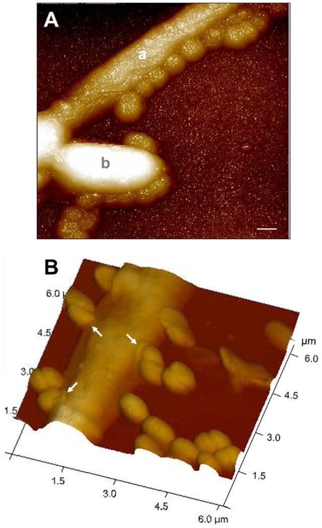
Fig. 3.
Scanning Probe Microscope (SPM) images of C. albicans SC5314 hypha-forming cells interacting with S. gordonii DL1. Filamentation-induced C. albicans cells in YPT-Glc medium were incubated with S. gordonii cells for 1 h at 37°C. Cells were then deposited onto glass cover slips and imaged in contact mode as described in Experimental procedures. Panel A, undried specimen showing hyphal filament (a) with smaller streptococcal cells attached along its length. A budding pseudohypha (b) appears also to have streptococci attached. Panel B, dried specimen showing numerous streptococcal cells in close physical contact with a C. albicans hyphal filament. At the point of contact there is an annular modification visible on the C. albicans cell surface (arrowed), implying a hyphal cell surface structural response. Note that quite often the streptococcal cell septum region was involved in binding hyphae (see also Fig. 2B). Scale bar 0.5 μm.

